

Tracking Macular Changes: How Retinal Imaging Protects Richmond Seniors Moving Into Fall
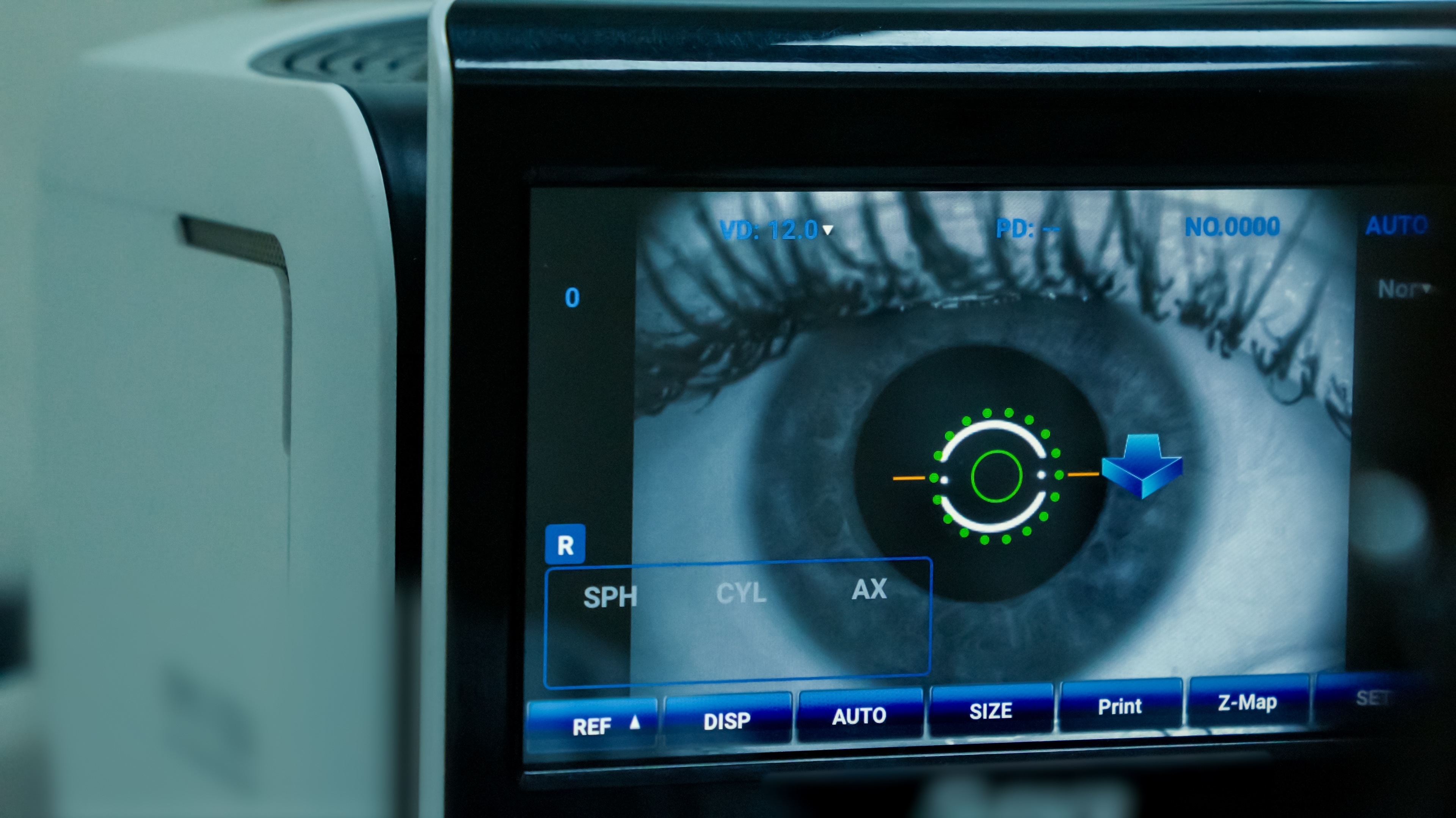
As fall approaches, many Richmond seniors use the change of season as a reminder to catch up on routine health appointments. Your eyes deserve a place on that list, especially because conditions affecting the macula can begin changing before you notice a major difference in everyday vision.
At Dr. Gina Dyda-Schmid Optometrist and Associates, we use advanced retinal imaging as part of comprehensive eye care to examine and document the health of the retina. These images can help us identify changes and compare findings from one visit to the next.
Why Does the Macula Matter?
The macula is a small area at the center of the retina responsible for detailed central vision. You rely on it when reading, driving, recognizing faces, and completing tasks that require fine visual detail.
Age-related macular degeneration, or AMD, affects this important area. Because early AMD may develop without obvious symptoms, clear vision today does not necessarily mean there are no changes occurring inside the eye.
Regular comprehensive eye exams become increasingly important with age, particularly for patients with additional risk factors such as a family history of macular degeneration or smoking history.
What Can Retinal Imaging Show?
Retinal imaging gives us a detailed view of structures at the back of the eye. At our Richmond office, we use Optomap technology to capture an ultra-widefield image of the retina.
Depending on your eye health, retinal imaging can help us document areas such as:
The macula and surrounding retinal tissue
The optic nerve
Retinal blood vessels
Areas of pigmentation or other retinal changes
Peripheral portions of the retina
Findings that may need additional evaluation or monitoring
The images become part of your eye health record, giving us a useful reference for future examinations.
Why Is Comparing Images Over Time Important?
One of the advantages of digital retinal imaging is the ability to compare today's findings with images from previous visits.
A small retinal change may be difficult for a patient to notice in daily life, but side-by-side images can help an eye doctor determine whether something appears stable or has changed over time. This is especially valuable when monitoring age-related conditions that may progress gradually.
Retinal imaging does not replace every other part of a comprehensive eye examination. Instead, it provides another layer of information that can help us decide whether additional testing, closer monitoring, or referral for treatment is appropriate.
What Changes Can Macular Degeneration Cause?
AMD primarily affects central rather than peripheral vision. As the condition progresses, patients may notice that fine details become more difficult to see.
Straight lines may begin appearing wavy, words may seem blurry, or a dark or empty area can develop near the center of vision. Recognizing faces may also become more difficult.
These symptoms should be evaluated promptly rather than waiting for your next routine appointment. Sudden or significant central vision changes can require timely assessment.
Does Retinal Imaging Mean You Do Not Need Dilation?
Not necessarily. Optomap provides a wide digital view of the retina, but retinal imaging and a dilated examination serve different purposes.
Depending on your age, symptoms, medical history, previous findings, and individual risk factors, your eye doctor may recommend imaging, dilation, or both. The goal is to use the appropriate tools to get the clearest possible picture of your retinal health.
Make Fall Eye Care Part of Healthy Aging
Routine eye care becomes particularly valuable as you get older because several eye conditions become more common with age. Tracking retinal changes over time can help identify concerns before they have a greater impact on vision and give you a clearer understanding of how your eyes are changing.
If it has been a while since your last comprehensive eye exam, schedule a visit with Dr. Gina Dyda-Schmid Optometrist and Associates at 11736 W Broad St #106B, Richmond, VA 23233 by calling or texting (804) 360-1590. Our team can evaluate your retinal health and use advanced imaging to help monitor macular changes as you move into fall.

All Eye
Care Services

Keep
In Touch


Let’s keep in touch!
Contact Us

Office Hours
- Monday 10:00am - 5:00pm
- Tuesday 10:00am - 6:00pm
- Wednesday 10:00am - 5:00pm
- Thursday 10:00am - 6:00pm
- Friday 10:00am - 5:00pm
- Saturday 10:00am - 4:00pm
- Sunday Closed
© 2026 Dr. Gina Dyda-Schmid Optometrist and Associates | All rights Reserved | Accessibility Statement | Privacy Policy | Sitemap
Powered by:
